Innovative Modification for Orthognathic Splint
Piyush Bolya*,1, Smarika Kothari2, Archana Nagora2, Vaibhav Jain3, Priyashree Joshi3, Snehal Dupare4
1Head and Professor, Department of Orthodontics and Dentofacial Orthopedics, Pacific Dental College and Research Center, Udaipur, India
2Senior Lecturer, Department of Orthodontics and Dentofacial Orthopedics, Pacific Dental College and Research Center, Udaipur, India
3Consultant orthodontist, India
4Reader, Department of Orthodontics and Dentofacial Orthopedics, Yogita Dental College and Hospital, Khed, India
*Corresponding Author: Piyush Bolya, Head and Professor, Department of Orthodontics and Dentofacial Orthopedics, Pacific Dental College and Research Center, Udaipur, India
Received: 21 May 2025; Accepted: 26 May 2022; Published: 30 May 2025
Article Information
Citation: Piyush Bolya, Smarika Kothari, Archana Nagora, Vaibhav Jain, Priyashree Joshi, Snehal Dupare. Innovative Modification for Orthognathic Splint. Journal of Environmental Science and Public Health. 9 (2025): 18-21.
View / Download Pdf Share at FacebookAbstract
Le Fort I osteotomy is a frequently performed surgical procedure in patients with cleft lip and palate, often complicated by maxillary hypoplasia and scarring from previous corrective surgeries. These factors can make maxillary mobilization challenging and negatively impact long-term stability. Acrylic occlusal wafers, commonly used in both cleft and non-cleft osteotomies, help establish proper jaw alignment, but cleft patients require significantly more force for mobilization. To address this, a modified acrylic occlusal splint was developed, incorporating metal loops for improved intermaxillary fixation. The construction process involves standard techniques, with additional steps to ensure optimal engagement and comfort. The modified splint offers several advantages, including increased surgical stability, cost-effectiveness, and protection of oral tissues, making it a valuable tool for cleft lip and palate osteotomies.
Keywords
Le Fort I osteotomy, cleft lip and palate, maxillary hypoplasia, acrylic occlusal splint, intermaxillary fixation, maxillary mobilization, cleft surgery, orthognathic surgery, jaw alignment, surgical innovation
Article Details
Introduction
Le Fort I osteotomy is one of the most frequently performed surgeries in patients with cleft lip and palate. In these cases, a hypoplastic maxilla is often observed, typically due to previous corrective procedures for the cleft, which can result in scarring and distortion of the soft tissues. The presence of scar tissue presents extra challenges during surgery, as it complicates the mobilization of the maxilla and can impact long-term stability. Acrylic occlusal wafers are frequently used in osteotomies for both cleft and non-cleft patients to establish the planned jaw relationship by properly positioning the mobilized maxilla. However, for cleft patients, mobilization demands significantly more force. To address this, we have altered the design of the occlusal splint, resulting in a new version. These slight modifications, combined with a low cost, offer several advantages.
Design
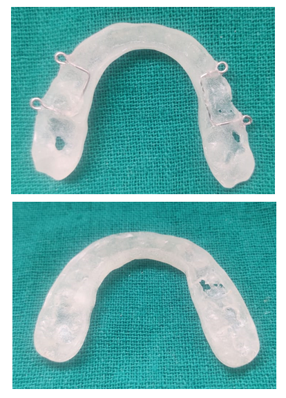
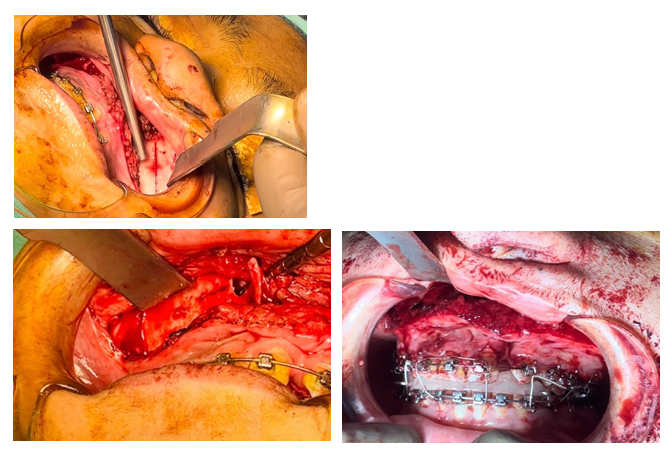
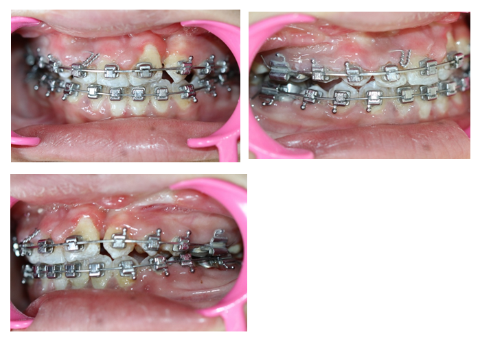
A standard acrylic occlusal splint offers occlusal coverage only. However, a modified version, made from the same material, not only provides occlusal coverage but also includes metal loops for proper intermaxillary fixation. The procedures involved are similar to those used in standard osteotomies, but this modified splint offers several advantages. By ensuring better engagement between the mandible and maxilla, it enhances stability, allowing for the application of greater force with more control. Additionally, this design helps protect the dentition, gingiva, and palatal soft tissues.
Construction
1.A separating medium is applied to the casts using a paintbrush.
2.Self-curing acrylic is shaped into a U-shaped configuration and placed on the lower occlusal surface.
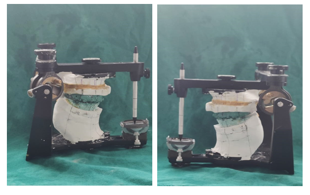
3.Metal loops are incorporated at least in five locations: between the central incisors, distal to the lateral incisors, and distal to the second premolars. The articulator is then closed.
4.While the acrylic is still in its soft (dough) stage, trim and contour any excess material on the labial, lingual, and buccal surfaces to prevent interference with the orthodontic appliance and to ensure that the acrylic does not tear during this process.
5.Complete routine finishing and polishing to ensure patient comfort and maintain proper oral hygiene.
Conclusion
The modified acrylic occlusal splint, designed for Le Fort I osteotomy in patients with cleft lip and palate, significantly enhances the stability and control of maxillary mobilization. By incorporating metal loops for improved intermaxillary fixation, this design allows for greater force application while protecting the dentition, gingiva, and palatal tissues. The modification offers several advantages over traditional splints, including better engagement between the mandible and maxilla, improved surgical outcomes, and cost-effectiveness. This innovative approach addresses the unique challenges presented by cleft patients, providing a more reliable and controlled means of achieving the planned jaw relationship. As such, the modified splint represents a valuable tool for improving the success and long-term stability of osteotomies in cleft lip and palate patients.
References
- Saltaji H, Major MP, Alfakir H, et al. Maxillary advancement with con-ventional orthognathic surgery in patients with cleft lip and palate: is it astable technique? J Oral Maxillofac Surg 70 (2012): 2859–66.
- Thongdee P, Samman N. Stability of maxillary surgical movement in uni-lateral cleft lip and palate with preceding alveolar bone grafting. CleftPalate Craniofac J 42 (2005): 664–74.
- Scolozzi P. Distraction osteogenesis in the management of severe max-illary hypoplasia in cleft lip and palate patients. J Craniofac Surg 19 (2008): 1199–214.
- Bamber MA, Harris M. The role of the occlusal wafer in orthognathicsurgery; a comparison of thick and thin intermediate osteotomy wafers. JCraniomaxillofac Surg 23 (1995): 396–400.
- Lindorf HH. Surgical-cephalometric bite reconstruction (double splint method). Dtsch Zahnarztl Z 32 (1977): 260-1.
- Luhr HG, Kubein-Meesenburg D, Schwestka-Polly R. The importance and technique of temporo-mandibular joint positioning in the sagittal splitting of the mandible. Fortschr Kieferorthop 52 (1991): 66-72.
- Leib AM. The occlusal bite splint—a non-invasive therapy for occlusal habits and temporo-mandibular disorders. Comp end Contin Educ Dent 11 (1996): 1081-4, 1086-88.


 Impact Factor: * 3.6
 Acceptance Rate: 76.49%
  Time to first decision: 10.4 days
  Time from article received to acceptance: 2-3 weeks